


Verified AB-Abdomen Exam Dumps Q&As - Provide AB-Abdomen with Correct Answers
Pass Your AB-Abdomen Dumps Free Latest ARDMS Practice Tests
ARDMS AB-Abdomen Exam Syllabus Topics:
| Topic | Details |
|---|---|
| Topic 1 |
|
| Topic 2 |
|
| Topic 3 |
|
| Topic 4 |
|
NEW QUESTION # 82
Which sonographic finding is most consistent with scrotal inflammation?
- A. Hyperemia
- B. Granuloma
- C. Hydrocele
- D. Abscess
Answer: A
Explanation:
Scrotal inflammation, such as epididymitis or orchitis, typically presents with increased blood flow (hyperemia) on color Doppler sonography. This finding reflects the inflammatory process and vascular dilation. Abscesses, granulomas, or hydroceles may be present but are not as consistent or specific for inflammation.
According to AIUM Practice Parameters and Rumack's Diagnostic Ultrasound:
"In acute inflammation, color Doppler ultrasound demonstrates prominent hyperemia of the epididymis or testis." Reference:
Rumack CM, Wilson SR, Charboneau JW, Levine D. Diagnostic Ultrasound. 5th ed. Elsevier, 2017.
AIUM Practice Parameter for Scrotal Ultrasound, 2020.
-
NEW QUESTION # 83
Which laboratory value stays elevated longest and is considered the most reliable in diagnosing pancreatitis?
- A. Amylase
- B. Somatostatin
- C. Trypsin
- D. Lipase
Answer: D
Explanation:
Lipase is the most sensitive and specific laboratory marker for diagnosing acute pancreatitis. It rises earlier, remains elevated longer (up to 14 days), and is more pancreas-specific than amylase. Amylase may normalize within 48-72 hours and may also be elevated in non-pancreatic conditions.
According to ACG (American College of Gastroenterology) Guidelines:
"Serum lipase is preferred over amylase due to its higher sensitivity, specificity, and prolonged elevation in pancreatitis." Reference:
American College of Gastroenterology (ACG) Clinical Guideline: Management of Acute Pancreatitis, 2013.
Rumack CM, Wilson SR, Charboneau JW, Levine D. Diagnostic Ultrasound. 5th ed. Elsevier, 2017.
-
NEW QUESTION # 84
Which type of artifact is indicated by the arrows on this image?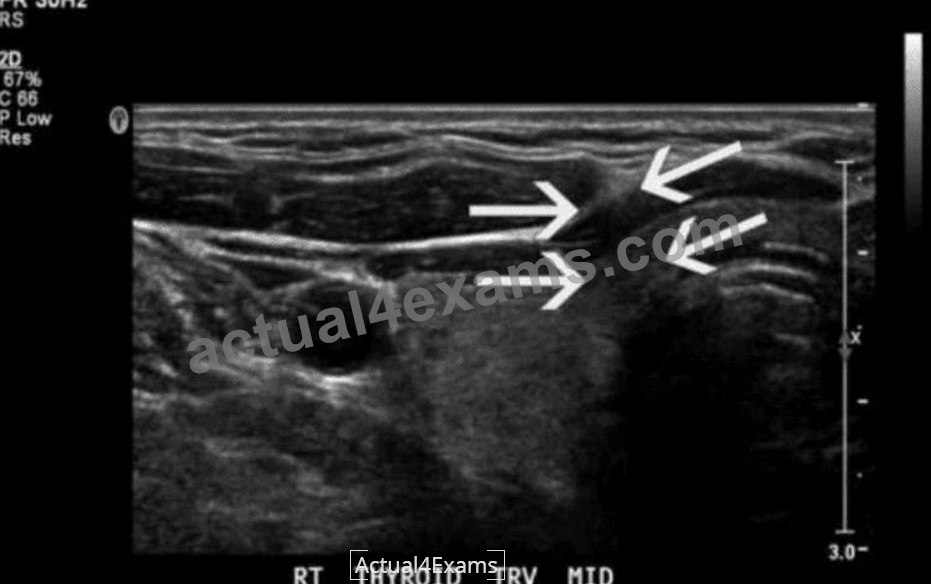
- A. Comet tail
- B. Edge shadow
- C. Focal enhancement
- D. Speed error
Answer: B
Explanation:
The ultrasound image of the thyroid clearly shows posterior shadowing originating from the lateral edges of a rounded structure, which is indicative of edge shadow artifact. Edge shadowing occurs when an ultrasound beam passes tangentially to a rounded or curved structure, such as a cyst or blood vessel. The difference in sound wave refraction and beam divergence at the edges leads to decreased echo signals deep to the edges, creating linear hypoechoic bands - which is exactly what the arrows are pointing to in the image.
Edge shadow artifact is purely a result of beam physics and not a real anatomic or pathologic finding.
Key characteristics of edge shadowing:
* Appears as a narrow, linear hypoechoic (dark) shadow extending deep to the edge of a curved interface (e.g., cyst, vessel, thyroid nodule)
* Caused by refraction and beam deflection, leading to reduced beam intensity distal to the edges
* Most commonly seen adjacent to cysts or fluid-filled structures
Differentiation from other options:
* A. Focal enhancement: Appears as increased echogenicity distal to a fluid-filled structure due to lower attenuation of the sound beam through fluid (opposite of shadowing).
* C. Speed error: A less common artifact that results in displacement of structures due to variation in assumed sound speed.
* D. Comet tail: A reverberation artifact that appears as a series of closely spaced bright echoes, often associated with metallic objects or cholesterol crystals in adenomyomatosis.
References:
Rumack CM, Wilson SR, Charboneau JW, Levine D. Diagnostic Ultrasound. 5th Edition. Elsevier, 2018.
Chapter: Ultrasound Physics and Artifacts, pp. 38-42.
Kremkau FW. Sonography Principles and Instruments. 9th Edition. Elsevier, 2015. Chapter: Image Artifacts, pp. 132-136.
NEW QUESTION # 85
Which retroperitoneal finding is most likely associated with trauma?
- A. Urinoma
- B. Fibrosis
- C. Adenoma
- D. Neuroblastoma
Answer: A
Explanation:
Urinomas are collections of urine in the retroperitoneum that result from trauma, surgery, or obstruction causing urine leakage. Trauma is a frequent cause of urinoma formation due to disruption of the urinary tract.
According to Rumack's Diagnostic Ultrasound:
"Urinomas may develop as a complication of trauma, surgery, or obstructive uropathy with urinary extravasation into the retroperitoneum." Reference:
Rumack CM, Wilson SR, Charboneau JW, Levine D. Diagnostic Ultrasound. 5th ed. Elsevier, 2017.
AIUM Practice Parameter for Renal Ultrasound, 2020.
-
NEW QUESTION # 86
Which congenital disorder is most consistent with the finding identified by the arrow on this image?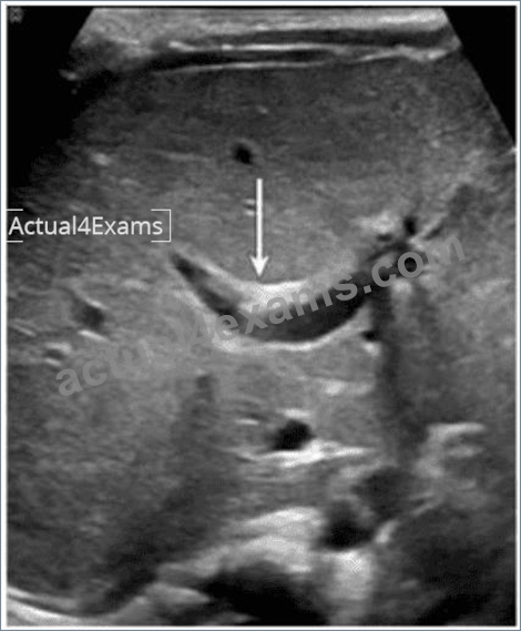
- A. Alagille syndrome
- B. Biliary atresia
- C. Caroli disease
- D. Sclerosing cholangitis
Answer: C
Explanation:
The image demonstrates a characteristic "central dot sign" - a hallmark finding of Caroli disease. This is best appreciated on ultrasound as a cystic dilation of the intrahepatic bile ducts with a central echogenic dot or linear structure (which corresponds to the portal vein and fibrous tissue within the dilated duct). The arrow in the image points to one such dilated duct.
Caroli disease is a rare congenital disorder characterized by segmental, saccular dilation of intrahepatic bile ducts. It is often associated with congenital hepatic fibrosis and may predispose to cholangitis, stone formation, and even cholangiocarcinoma.
Key ultrasound features of Caroli disease:
* Cystic or saccular dilations of the intrahepatic bile ducts
* The "central dot sign" - echogenic focus in the center of the dilated ducts (representing portal vein radicle or fibrous tissue)
* May show associated hepatosplenomegaly or signs of portal hypertension Differentiation from other options:
* A. Sclerosing cholangitis: Typically causes diffuse or segmental biliary ductal wall thickening and stricturing; does not present with cystic dilations.
* B. Alagille syndrome: A multisystem disorder often characterized by a paucity of intrahepatic bile ducts, not dilation.
* D. Biliary atresia: Presents in infancy with obliteration of extrahepatic bile ducts, echogenic "triangular cord" sign, and absence of a visible gallbladder. It does not cause ductal dilation.
References:
Rumack CM, Wilson SR, Charboneau JW, Levine D. Diagnostic Ultrasound. 5th Edition. Elsevier, 2018.
Chapter: Biliary System, pp. 152-155.
Radiopaedia.org. Caroli disease. https://radiopaedia.org/articles/caroli-disease American College of Radiology (ACR). ACR-SPR Practice Parameter for the Performance of Pediatric Abdominal Ultrasound, 2022.
NEW QUESTION # 87
Which imaging technique best demonstrates ureteral patency?
- A. Graded compression
- B. Spectral Doppler
- C. Color Doppler
- D. Gray scale
Answer: C
Explanation:
Color Doppler imaging can detect ureteral jets, which represent urine entering the bladder from the ureters.
The presence of bilateral ureteral jets confirms ureteral patency. Gray scale may show hydronephrosis but does not directly assess flow.
According to Rumack's Diagnostic Ultrasound:
"Color Doppler demonstrates ureteral jets within the bladder, confirming ureteral patency." Reference:
Rumack CM, Wilson SR, Charboneau JW, Levine D. Diagnostic Ultrasound. 5th ed. Elsevier, 2017.
AIUM Practice Parameter for Renal Ultrasound, 2020.
-
NEW QUESTION # 88
Which sonographic finding is most consistent with this image of the abdominal aorta?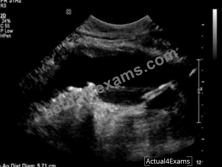
- A. Stenosis
- B. Aneurysm
- C. Occlusion
- D. Dissection
Answer: B
Explanation:
The ultrasound image provided shows a transverse view of the abdominal aorta, with a clearly measured aortic diameter of 5.71 cm. A normal adult abdominal aorta should measure less than 3.0 cm in anterior- posterior diameter. Any measurement exceeding this threshold is defined as an abdominal aortic aneurysm (AAA).
In this case, the dilation is well beyond the 3.0 cm threshold, confirming the presence of an aneurysm. The rounded, anechoic/heterogeneous central lumen surrounded by echogenic arterial wall layers further supports this diagnosis.
Comparison of answer choices:
* A. Stenosis: Would show a narrowed lumen with turbulent, aliasing flow on Doppler, not a dilated aorta.
* B. Dissection: Typically shows an echogenic intimal flap separating true and false lumens; no flap is visible here.
* C. Aneurysm: Correct. The aorta's transverse diameter (5.71 cm) confirms the presence of an aneurysm.
* D. Occlusion: Would appear as a lack of flow with thrombus or echogenic content filling the lumen, not dilation.
References:
Rumack CM, Wilson SR, Charboneau JW, Levine D. Diagnostic Ultrasound, 5th ed. Elsevier; 2017.
Society for Vascular Surgery Guidelines: Management of Abdominal Aortic Aneurysms (Chaikof et al., J Vasc Surg, 2018).
Hagen-Ansert SL. Textbook of Diagnostic Sonography, 8th ed. Elsevier; 2017.
NEW QUESTION # 89
Which abnormality is the most common adult adrenal tumor?
- A. Pheochromocytoma
- B. Adenoma
- C. Neuroblastoma
- D. Adrenal cortical carcinoma
Answer: B
Explanation:
Adrenal adenomas are the most common adrenal tumors in adults. They are often discovered incidentally (adrenal incidentalomas) and are usually nonfunctioning, though some may secrete cortisol or aldosterone.
Neuroblastoma is common in children, pheochromocytomas are rarer catecholamine-producing tumors, and adrenal cortical carcinoma is malignant but much less common than adenomas.
According to Rumack's Diagnostic Ultrasound:
"Adrenal adenomas are the most common adrenal masses in adults, frequently identified incidentally on imaging studies." Reference:
Rumack CM, Wilson SR, Charboneau JW, Levine D. Diagnostic Ultrasound. 5th ed. Elsevier, 2017.
ACR Incidental Findings Committee Guidelines for Adrenal Masses, 2017.
-
NEW QUESTION # 90
Which vessel is located directly proximal to the origination of the renal arteries?
- A. Splenic vein
- B. Hepatic artery
- C. Left portal vein
- D. Superior mesenteric artery
Answer: D
Explanation:
The renal arteries originate from the abdominal aorta just inferior to the superior mesenteric artery (SMA).
The SMA arises anteriorly from the abdominal aorta at the level of L1, and just below it, the renal arteries branch laterally. The splenic vein, portal vein, and hepatic artery are located more superiorly in relation to the renal arteries.
According to Moore's Clinically Oriented Anatomy:
"The superior mesenteric artery arises from the anterior surface of the abdominal aorta just above the renal arteries." (Moore KL et al., Clinically Oriented Anatomy, 8th ed.) Reference:
Moore KL, Dalley AF, Agur AMR. Clinically Oriented Anatomy. 8th ed. Wolters Kluwer, 2018.
Gray's Anatomy for Students, 4th ed., Elsevier, 2019.
NEW QUESTION # 91
Which condition is most likely to develop after splenic trauma?
- A. Splenule
- B. Splenosis
- C. Target lesion
- D. Hamartoma
Answer: B
Explanation:
Splenosis refers to autotransplantation of splenic tissue following splenic trauma or splenectomy. After rupture, splenic fragments may implant throughout the peritoneal cavity and vascularize, forming multiple nodules of functional splenic tissue. Splenosis is typically asymptomatic and discovered incidentally on imaging.
Splenule (A) is a congenital accessory spleen, not related to trauma.
Hamartoma (C) is a benign primary splenic lesion.
Target lesion (D) generally refers to sonographic appearance seen in metastases or infections, not post-trauma.
Reference Extracts:
Mortele KJ, et al. "Multimodality imaging of splenic lesions and the spleen." Radiographics. 2004;24(4):1137-
1163.
Rumack CM, Wilson SR, Charboneau JW, Levine D. Diagnostic Ultrasound. 5th ed. Elsevier, 2017.
-
NEW QUESTION # 92
Which sonographic finding is associated with normal postprocedural Doppler of a transjugular intrahepatic portosystemic shunt (TIPS)?
- A. Hepatofugal flow in the intrahepatic portal venous branches
- B. Hepatopetal flow in the intrahepatic portal venous branches
- C. Low-velocity flow throughout stent
- D. Triphasic flow throughout stent
Answer: B
Explanation:
After successful TIPS placement, the intrahepatic portal venous branches continue to exhibit hepatopetal (toward the liver) flow, while the stent itself shows continuous, relatively high-velocity monophasic flow.
Hepatofugal flow in intrahepatic branches may indicate shunt dysfunction.
According to Zwiebel's Introduction to Vascular Ultrasound:
"Normal post-TIPS Doppler shows hepatopetal flow in the intrahepatic portal veins and continuous high- velocity flow within the stent." Reference:
Zwiebel WJ, Pellerito JS. Introduction to Vascular Ultrasound. 6th ed. Elsevier, 2019.
AIUM Practice Parameter for the Performance of Portal Venous Ultrasound, 2020.
-
NEW QUESTION # 93
Which structure is indicated by the arrow in this image?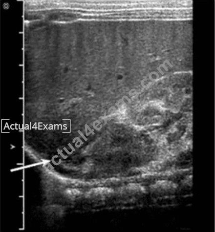
- A. Diaphragm
- B. Bowel loop
- C. Kidney
- D. Adrenal gland
Answer: D
Explanation:
The structure indicated by the arrow in the ultrasound image is the adrenal gland. On ultrasound, the adrenal gland in neonates and infants is relatively large and has a distinctive "Y" or "V" shape in the transverse view.
It is located superior and slightly medial to the upper pole of the kidney.
In this image, the arrow is pointing to a hypoechoic, curved structure with a thin echogenic central stripe, representing the fetal adrenal gland. This echogenic stripe corresponds to the adrenal medulla, while the surrounding hypoechoic area represents the cortex.
Differential features:
* A. Kidney: While the kidney is visualized posterior to the adrenal gland and shows a reniform shape with a central echogenic sinus and peripheral cortex, it is not the structure being directly pointed to by the arrow.
* B. Bowel loop: Bowel has variable echogenicity with peristalsis and shadowing from air. It does not have the consistent morphology or location seen in the image.
* C. Diaphragm: Appears as a thin, hyperechoic linear structure separating the thoracic cavity from the abdomen. It is seen more superiorly than the indicated structure and lacks the "Y" or "V" adrenal configuration.
Key Anatomical Landmarks:
* The adrenal glands are located in the retroperitoneum, superior to the kidneys, and appear prominent on ultrasound in neonates.
* In transverse view, the right adrenal gland is anterior to the crus of the diaphragm and posterior to the inferior vena cava (IVC).
References:
Rumack CM, Wilson SR, Charboneau JW, Levine D. Diagnostic Ultrasound. 5th Edition. Elsevier, 2018.
Chapter: Adrenal Glands and Retroperitoneum, pp. 291-295.
American Institute of Ultrasound in Medicine (AIUM) Practice Parameter for the Performance of an Ultrasound Examination of the Abdomen and/or Retroperitoneum. 2020.
NEW QUESTION # 94
Which technique is used to demonstrate the finding in this video?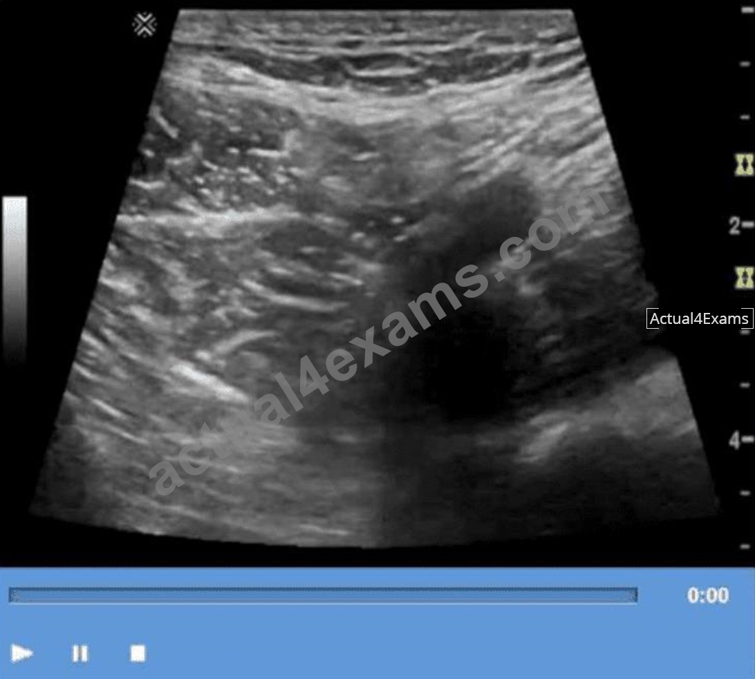
- A. Deep inspiration
- B. Compression
- C. Exhalation
- D. Valsalva
Answer: B
Explanation:
The technique shown in the video is compression. In ultrasound imaging-especially of soft tissue masses, the bowel, or venous structures-compression is used to evaluate the compressibility of structures. The image demonstrates a classic grayscale ultrasound view of a lesion or structure being compressed with the probe.
Compression sonography is particularly important in:
* Evaluating venous patency (e.g., for deep vein thrombosis)
* Differentiating cystic from solid structures
* Evaluating bowel wall abnormalities or intussusception
* Assessing lymph nodes and soft tissue masses (as shown here)
When a structure compresses easily under probe pressure, it suggests that the lesion is fluid-filled or soft. In contrast, incompressibility may indicate a solid mass or thrombus.
Differentiation from other options:
* B. Valsalva: Involves forced expiration against a closed airway, used primarily to assess venous reflux or inguinal hernias-not what is demonstrated here.
* C. Exhalation: A respiratory maneuver that passively alters thoracoabdominal pressure, not actively performed by the operator or causing focal structural change.
* D. Deep inspiration: Used to improve visualization of the liver, diaphragm, or gallbladder-not to evaluate the compressibility of soft tissue.
References:
Rumack CM, Wilson SR, Charboneau JW, Levine D. Diagnostic Ultrasound. 5th Edition. Elsevier, 2018.
Chapter: Ultrasound Technique and Physics, pp. 35-39.
AIUM Practice Parameter for the Performance of a Diagnostic Ultrasound Examination, 2020.
NEW QUESTION # 95
Which condition is most consistent with the findings in the image below?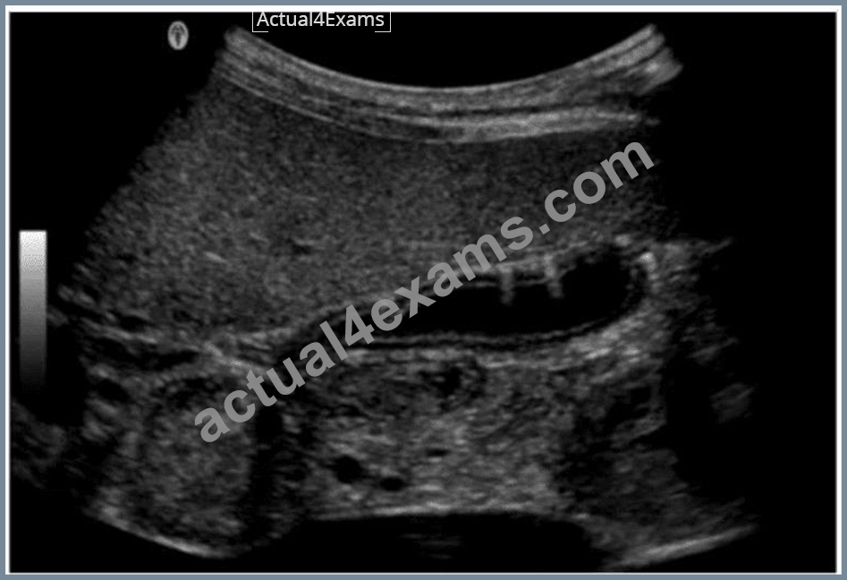
- A. Porcelain gallbladder
- B. Emphysematous cholecystitis
- C. Adenomyomatosis
- D. Gangrenous cholecystitis
Answer: B
Explanation:
The ultrasound image shows echogenic foci with dirty shadowing and reverberation artifacts within the gallbladder wall and lumen. These features are characteristic of emphysematous cholecystitis, a severe, life- threatening variant of acute cholecystitis caused by gas-forming organisms (e.g., Clostridium or E. coli) infecting the gallbladder wall.
Sonographic features of emphysematous cholecystitis:
* Echogenic gas within the gallbladder wall or lumen
* Reverberation or "dirty" shadowing artifacts
* May show intramural gas bubbles or "ring-down" artifact
* Often seen in diabetic or immunocompromised patients
* No gallstones may be present ("acalculous cholecystitis")
Clinical context:
* More common in elderly men and diabetics
* Presents with right upper quadrant pain, fever, and leukocytosis
* Surgical emergency due to risk of perforation and sepsis
Differentiation from other options:
* A. Adenomyomatosis: Involves gallbladder wall thickening with "comet tail" artifacts due to Rokitansky-Aschoff sinuses, not intramural gas.
* B. Porcelain gallbladder: Shows curvilinear calcification of the gallbladder wall - dense echogenic rim with posterior shadowing.
* C. Gangrenous cholecystitis: May show wall irregularity, intraluminal membranes, and absence of Doppler flow but lacks intramural gas.
References:
Rumack CM, Wilson SR, Charboneau JW, Levine D. Diagnostic Ultrasound. 5th Edition. Elsevier, 2018.
Chapter: Gallbladder and Biliary System, pp. 155-160.
American College of Radiology (ACR). Appropriateness Criteria for Right Upper Quadrant Pain, 2022.
Radiopaedia.org. Emphysematous cholecystitis: https://radiopaedia.org/articles/emphysematous-cholecystitis
NEW QUESTION # 96
Which exam type would be most beneficial for evaluating an elderly male patient who is complaining of dysuria and nocturia?
- A. Penile Doppler
- B. Renal Doppler
- C. Scrotal
- D. Pelvis limited
Answer: D
Explanation:
In elderly men, dysuria and nocturia often suggest lower urinary tract symptoms such as benign prostatic hyperplasia (BPH). A pelvis limited ultrasound is most appropriate to evaluate the bladder and prostate for enlargement, residual urine volume, or other causes of obstructive symptoms.
According to AIUM Practice Guidelines:
"Pelvic ultrasound can evaluate bladder volume, post-void residual, and prostate size in men with lower urinary tract symptoms." Reference:
AIUM Practice Parameter for the Performance of Ultrasound of the Prostate, 2020.
Rumack CM, Diagnostic Ultrasound, 5th ed. Elsevier, 2017.
-
NEW QUESTION # 97
Which thyroid condition is most likely caused by a viral infection?
- A. Graves
- B. Hashimoto
- C. De Quervain
- D. Abscess
Answer: C
Explanation:
De Quervain thyroiditis (subacute granulomatous thyroiditis) is often triggered by a viral infection. Patients may present with painful thyroid enlargement, elevated inflammatory markers, and transient hyperthyroidism.
Hashimoto's and Graves' diseases are autoimmune in nature.
According to Braverman's The Thyroid:
"Subacute (De Quervain) thyroiditis typically follows a viral upper respiratory tract infection and is characterized by thyroid pain and transient thyrotoxicosis." Reference:
Braverman LE, Cooper DS. The Thyroid: A Fundamental and Clinical Text. 11th ed. Wolters Kluwer, 2021.
American Thyroid Association Guidelines, 2016.
NEW QUESTION # 98
What is a major advantage of power Doppler over color flow Doppler?
- A. Decreased sensitivity to motion artifacts
- B. Doppler angle independent
- C. Ease of determining flow direction
- D. Improved signal-to-noise ratio
Answer: D
Explanation:
Power Doppler measures the amplitude (strength) of Doppler signals rather than frequency shift, making it more sensitive to low-velocity and small-vessel blood flow. Its primary advantage is an improved signal-to- noise ratio, allowing for better visualization of slow or weak flow.
* A: Power Doppler is more sensitive to motion artifacts, not less.
* B: It is still angle dependent, though somewhat less so than color Doppler.
* D: Power Doppler does not display flow direction (a limitation).
Reference Extracts:
* Kremkau FW. Sonography Principles and Instruments. 9th ed. Elsevier, 2015.
* Rumack CM, Wilson SR, Charboneau JW, Levine D. Diagnostic Ultrasound. 5th ed. Elsevier, 2017.
NEW QUESTION # 99
Which condition is most consistent with the sonographic appearance in this image of the abdominal wall?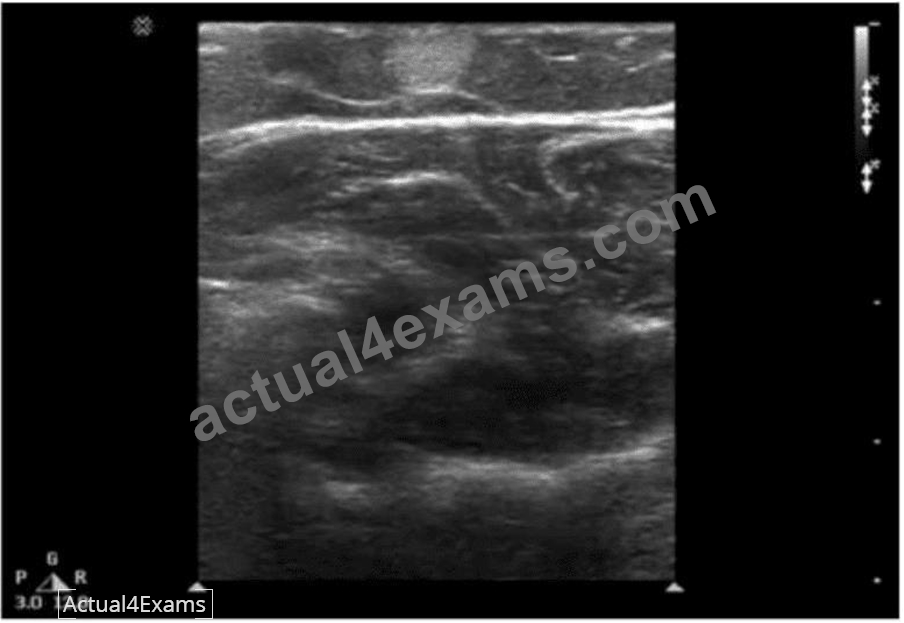
- A. Desmoid
- B. Metastasis
- C. Lipoma
- D. Fibroma
Answer: C
Explanation:
The ultrasound image demonstrates a well-defined, ovoid, hypoechoic to isoechoic mass within the subcutaneous tissue of the abdominal wall. The lesion appears compressible and shows linear striations parallel to the skin surface - a classic appearance of a lipoma.
Lipomas are the most common benign soft tissue tumors and frequently arise in the subcutaneous tissue. They are composed of mature adipose tissue and are typically asymptomatic unless large or compressing adjacent structures.
Sonographic features of a lipoma:
* Isoechoic to mildly hyperechoic or hypoechoic relative to subcutaneous fat
* Oval or elliptical in shape with well-defined margins
* Internal linear striations or "feathered" echotexture
* Compressible and non-vascular on Doppler imaging
* Located in subcutaneous fat plane parallel to the skin surface
Differentiation from other options:
* B. Fibroma: Typically appears as a homogeneous, hypoechoic mass but is far less common in the abdominal wall.
* C. Desmoid: Appears as an ill-defined or infiltrative mass within deeper soft tissues; more heterogeneous and may distort surrounding tissue planes.
* D. Metastasis: Often more irregular, heterogeneous, and may show increased vascularity or invasion into adjacent structures.
References:
Rumack CM, Wilson SR, Charboneau JW, Levine D. Diagnostic Ultrasound. 5th Edition. Elsevier, 2018.
Chapter: Musculoskeletal and Soft Tissue Ultrasound, pp. 1448-1450.
American Institute of Ultrasound in Medicine (AIUM) Practice Parameter for the Performance of a Diagnostic Ultrasound Examination of Soft Tissue Structures, 2020.
Radiopaedia.org. Lipoma (ultrasound): https://radiopaedia.org/articles/lipoma-ultrasound
NEW QUESTION # 100
......
Get Top-Rated ARDMS AB-Abdomen Exam Dumps Now: https://www.actual4exams.com/AB-Abdomen-valid-dump.html
AB-Abdomen Exam Dumps Pass with Updated Tests Dumps: https://drive.google.com/open?id=1oInrK-hmYAgr1-LTtuYQk-_BsisfKOBE